Introduces
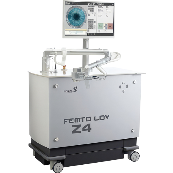
Z-LASIK
the most advanced All-Laser LASIK technology.
The Ziemer Z4 femtosecond laser empowers a 3D optimized Bladeless procedure.
3D Bladeless Topography-Guided LASIK
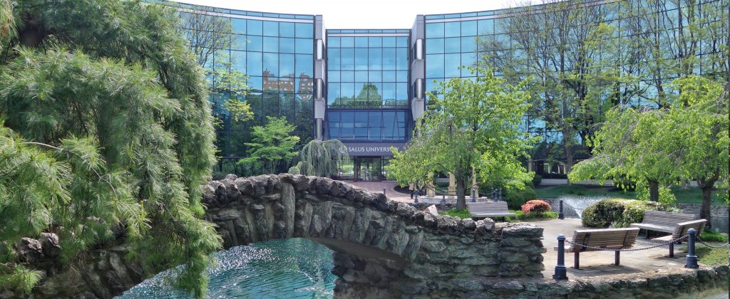
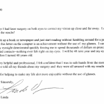
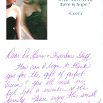
LewisLASIK at $5000 total for both eyes is second to none in quality, safety, experience, and clinical results. James S. Lewis, MD is a fellowship trained cornea surgical specialist, a Wills Eye Network Surgeon, and the Director of Refractive Surgery at the Pennsylvania College of Optometry (Salus University), the second largest training facility for eye doctors in the country. He uses the latest topographically-guided lasers and lectures nationally and internationally on surgical technique. Dr. Lewis has performed over 10,000 successful LASIK surgeries as well as over 100,000 major intra-ocular surgical procedures in the last three decades. He performs all LASIK surgery at LewisLASIK and always includes one year of free post-operative care.
Corporate LASIK Centers promising $299 an eye for LASIK are using a bait-and-switch technique to lure patients into their facility. Sales people then explain why you should pay for an upgraded laser, an upgraded post-operative program or “enhancements”. Often they simply claim you don’t qualify for the advertised low price. The average cost of LASIK in these centers is $6000. When deals seem “to good to be true” you can bet they are not true.
Don’t get upsold. Experience the most advanced LASIK procedure.
Click to Call Us
Directions to Our Offices
Schedule a Free Eval
LewisLASIK is a premier refractive surgery center. Our free LASIK evaluations are complete ocular examinations by experienced specialists. We emphasize customized results and individualized patient care. We are a private practice setting driven by excellence in clinical results.
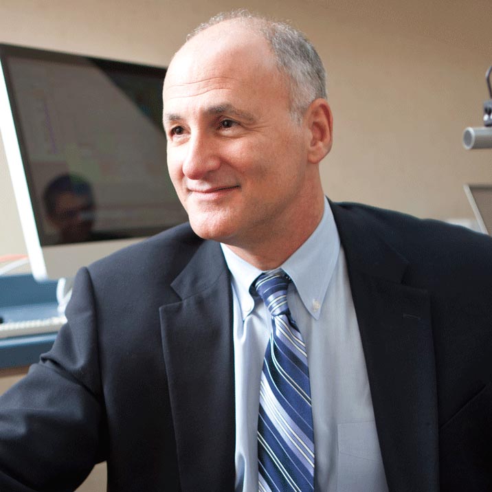
Dr. Lewis
James S. Lewis, MD is a graduate of Princeton University and Thomas Jefferson Medical College where he won awards in Ophthalmology and Research. He served as chief resident during his third year at Duke University Eye Center where he was offered a faculty position. Instead he travelled to South Australia for subspecialty training in Corneal and Refractive Surgery. He returned in 1990 and became Director of Cornea and Anterior Segment Surgery at Hahnemann University, in Philadelphia. He also served as Corneal Consultant to the Pennsylvania College of Optometry (now Salus University).
Advantages of Z-LASIK
LewisLASIK introduces Z-LASIK, the most advanced All-Laser LASIK technology. The Ziemer Z4 femtosecond laser empowers a 3D optimized Bladeless procedure.
- Increased comfort and safety
- Ultra precise technology
- Fast visual recovery
- Short procedure time
- Superior vision results immediately
Nidek Upgrades Custom LASIK
Better Than 20/20 &
Enhanced Night Driving Vision

Financing with CareCredit
6 Months:
No Interest
No Penalty
No Downpayment
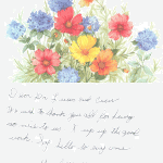
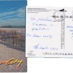
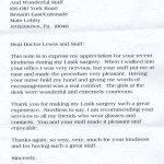
Dina

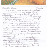
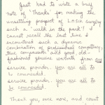
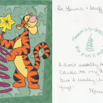
TJ Kelly
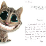
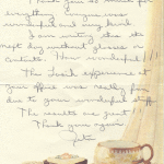
Paul
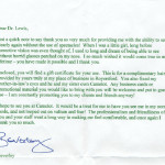
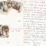
Erika
Adam G.
Patrick
Tony
Noreen
Jim & Betty
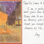
Amy

Steve K.

Stephanie
Karen
Jacqueline
Alan & Ann

Teo N.
Bertha
Peggy
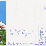
Beverley
Pat
Ed
Miriam
Susan
The senile one
Scott
Michael M.
Lilian
Fred
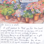
Ross
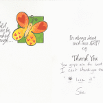
Sue
Lisa
Connie
Kelly
Suzanne
Kim
Kelly
Jimmy
Mrs. Anna
Myra
Jamie
Peggy
Deb
Linda
Marianne
Rebecca
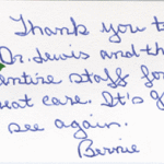
Bernie
Tracie
Dana
Delores
Dorothy
Harvey
Tore
Phyllis
Anna
Betty
William
Glorya
Millie & Tom
Dat
Joe
John
Patti
Marie
Rita
Karen
anonymous
Thelms
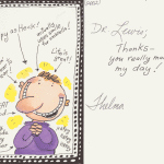
Elaine
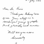
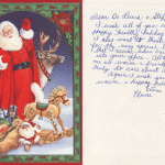
Sue

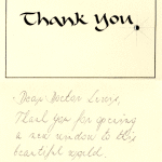
JGRW
Francis
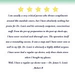
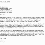
Robert R.
Anna
Kelly Ann
Donna
Paul
Steve
Joan
anonymous
Leonard, Herb & Bette
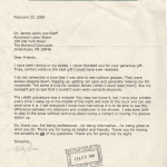
Jim F.
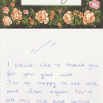
Nancy
H
V

Kathy
Paul
Mark
Thelma
Jim S.
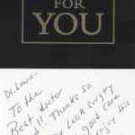
Leslie
Thomas
Teo
Dan
Nicole
Tom
Angela
Herta
Leslie
One Price for ALL
- Z-LASIK Technology
- NIDEK Custom LASIK
- Surgeon James S. Lewis, MD
LewisLASIK is a premier refractive surgery practice without a premium price. Our free LASIK evaluations are complete ocular examinations by experienced specialists. We emphasize customized results and individualized patient care.
Call for a consultation
215-886-9090LASIK TECHNOLOGY LEADER in the Delaware Valley